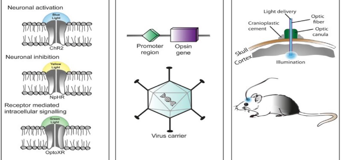
Optogenetics (from Greek, Modern optikós, meaning 'seen, visible') is a biological technique that involves the use of light to control cells in living tissue, typically neurons, that have been genetically modified to express light-sensitive ion channels. It is a neuromodulation method that uses a combination of techniques from optics and genetics to control and monitor the activities of individual neurons in living tissue—even within freely-moving animals—and to precisely measure these manipulation effects in real-time. The key reagents used in optogenetics are light-sensitive proteins. Neuronal control is achieved using optogenetic actuators like channelrhodopsin, halorhodopsin, and archaerhodopsin, while optical recording of neuronal activities can be made with the help of optogenetic sensors for calcium (GCaMP), vesicular release (synapto-pHluorin), neurotransmitter (GluSnFRs), or membrane voltage (arc lightning, ASAP1). Control (or recording) of activity is restricted to genetically defined neurons and performed in a spatiotemporal-specific manner by light. Source: https://en.wikipedia.org/wiki/Optogenetics
In 2010, optogenetics was chosen as the "Method of the Year" across all fields of science and engineering by the interdisciplinary research journal Nature Methods. At the same time, optogenetics was highlighted in the article on "Breakthroughs of the Decade" in the academic research journal Science. These journals also referenced recent public-access general-interest video Method of the year video and textual SciAm summaries of optogenetics.
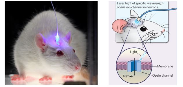
说到光遗传学(Optogenetics),很多英文文献的简要解释就是Controlling the Brain with Light,即通过光来控制大脑。字面理解起来很恐怖,马上联想到影帝小李子的《盗梦空间》,心有余悸啊。不过对于很多熟悉光遗传学技术的同学,脑海中肯定会浮现出以下这幅图,这是很多在体光遗传实验最常见的技术。在体光遗传技术更多的是采用光纤技术、将光源(LED或激光)引入小鼠脑区中,配合行为学研究或者电信号的记录。为什么光可以控制大脑呢?原来某些特定波长的光(如蓝光)可以打开大脑中神经细胞膜上的离子通道,使得这些神经细胞被动地变成兴奋状态,从而影响动物的某些行为和表现。这个对光敏感的离子通道(ChR2)属于一类独特的视蛋白,在蓝色光(中心波长470nm)照射的情况下,这些ChR2通道会打开,钙离子钠离子等会进入神经细胞内,引起细胞去极化产生动作电位。
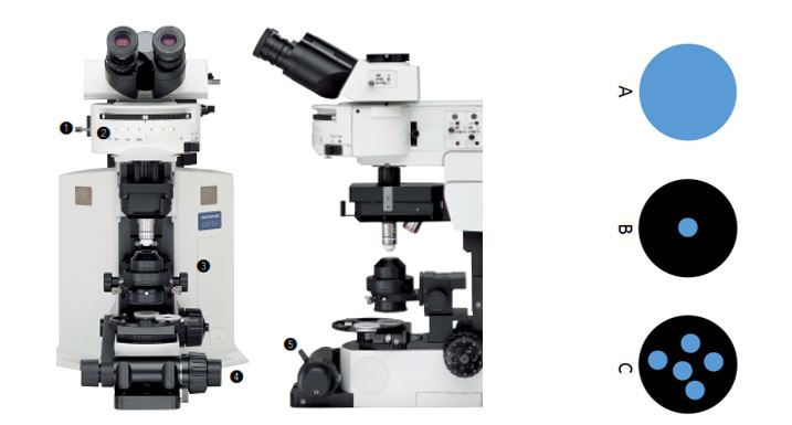
以上图片描述的在体光遗传学技术的优势是设备简单、价格亲民,但局限是光纤属于盲插,光纤顶端照明的具体位置并不能精确到单个细胞的级别。对于空间精度要求比较高的光遗传学实验,越来越多课题组采用显微镜作为辅助工具,将蓝光(LED或激光)精准地并且有一定频率地照射在视野内的一群或者指定的神经细胞上。下面三幅示意图中,黑色圆圈表示整个视野,蓝色小圆圈表示蓝光照射的区域:图A是整个视野都被照射(即视野内的所有神经元都被激活);图B是只有视野中心的区域被激活;图C是视野中多个区域有选择性地被照射。满足各种光遗传学实验的需求。 那么,如果在一台已有的电生理膜片钳显微镜上,是否可以方便快捷地实现上图中三种不同的显微镜上的光遗传学技术呢?答案是肯定的。采用不同类型的LED光源或者激光器,现在已经比较成熟的解决方案,满足这些对于空间精度要求高的实验。
 方案A) 采用全光谱白光LED荧光光源
传统电生理显微镜上的荧光光源采用的是汞灯,优势是光强较高,不足是汞灯灯泡寿命很短需经常更换(在屏蔽笼里更换调节很不方便),而且汞灯不能高频的开关。目前市面上的LED荧光光源稳定性及光强都能满足荧光显微镜的需求,并且LED光源可以通过外部TTL信号触发按照一定频率开关,通过放大器实现和电信号记录的完全同步。选择白光LED荧光光源需要注意以下几点:
1) 光源主机和显微镜分离,LED通过液芯光纤耦合到显微镜上,防止任何可能的电子噪声对于电信号记录的影响。这种方式也能防止光源的冷却模块产生的震动对于显微镜系统的影响。
2) LED调光旋钮可与光源主机分离,方便在屏蔽笼内直接手动调节LED光强。也可以通过USB线用软件来调节LED光强。
3) 宽光谱白光LED光源,这样可以直接替代汞灯光源,在显微镜上观察从紫外到近红外的荧光标记。同时,通过蓝色滤光片可将蓝光直接用于激活ChR2离子通道。
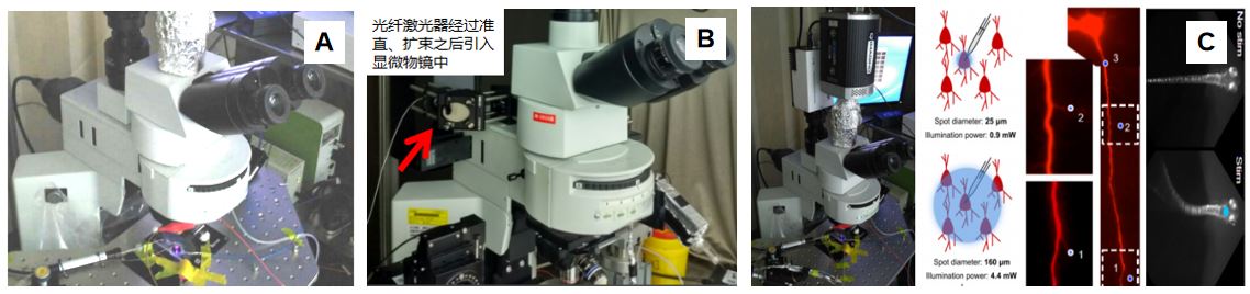 方案B) 采用外部光纤耦合激光器
将蓝光激光引入显微镜中,需要搭建光路、滤光片等配合,以保证激光通过物镜聚焦之后的光斑在视野中心,同时光斑大小后续可以调节。采用这个方案需要注意以下几点:
1) 激光的功率及功率稳定性都要有保证
2) 激光准直扩束之后的光斑大小需与物镜匹配
3) 选择合适的滤光片用于耦合激光
方案C) 采用DMD/DLP芯片照射多个区域
对于视野中多个区域同时照射的需求,基于数字微镜阵列技术的DMD/DLP芯片可以发挥很大的作用。类似在投影仪当中的应用,DLP芯片可以将蓝色或者黄色LED光源通过显微镜投射到样品上,单个或多个照明区域则可以通过软件设置。通过这个技术,在40倍物镜下最小照明区域可以达到0.4 x 0.4 um,实现单细胞、多细胞甚至细胞内的光刺激。采用这个方案需要注意以下几点:
1) 投影照明区域的平场均匀性,与光学成像区域匹配
2) 蓝光和黄光LED光源通过DLP芯片之后的功率
3) 投影芯片的刷新频率>1kHz,投影仪只有60Hz
4) 光刺激设备与电生理设备的同步
The
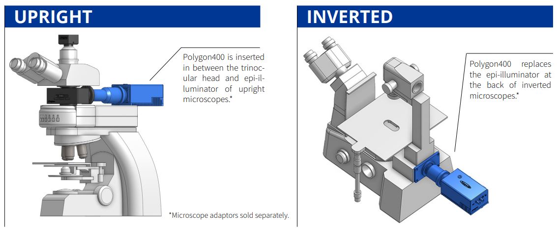
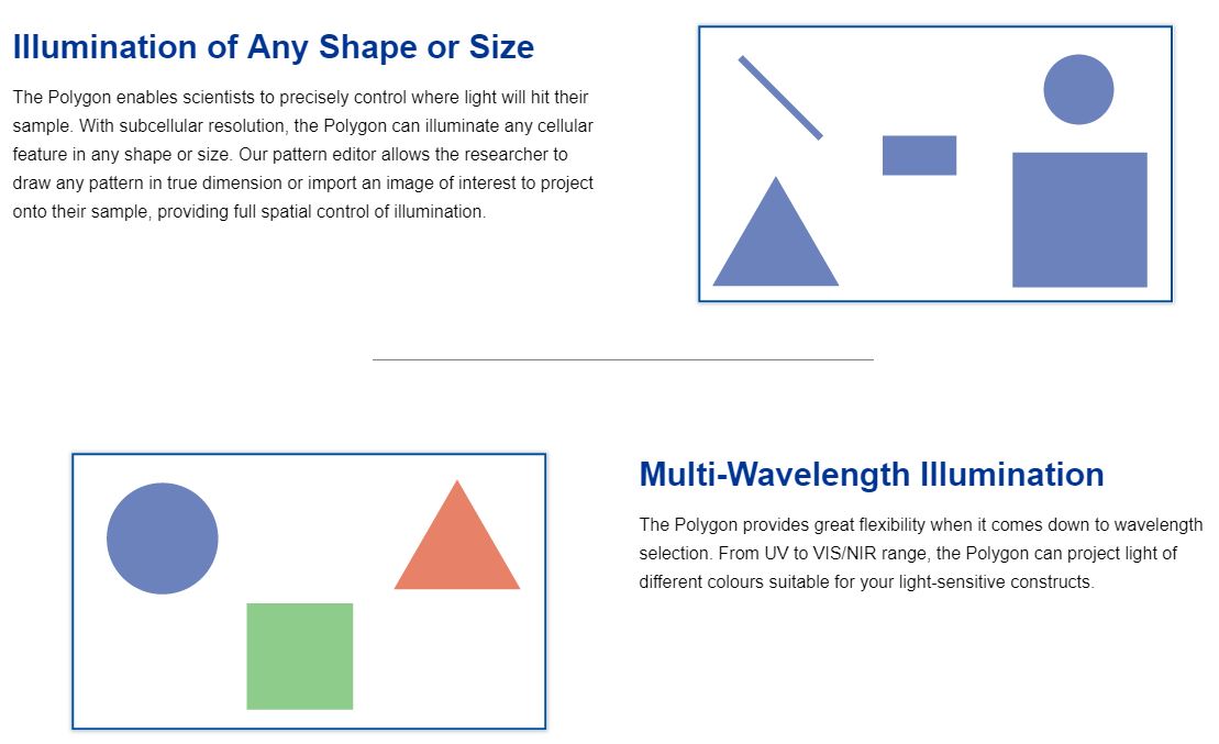
Polygon400 is Mightex’s market-leading pattern illuminator. It provides precise spatio-temporal control of light with single-cell or subcellular resolution, making it the perfect illumination tool for life science research. Compatible with any upright or inverted microscope, Polygon400 enables researchers to send light to anywhere on their specimen, and in any shape, size and complexity. In addition, multiple regions of interest can be illuminated simultaneously, and different wavelengths of light can be used with the Polygon400 for different bioscience applications. Furthermore, the Polygon400 can be seamlessly integrated via TTL into a larger system with other equipment such as electrophysiology tools or cameras.
 State-of-the-Art DMD Technology
The Polygon uses digital mirror device (DMD) technology to illuminate multiple regions simultaneously. A DMD is composed of hundreds of thousands of micro-mirrors that can be individually turned on to reflect light onto the sample. Thus, you can control each mirror to control the area(s) of illumination and create any number of different sized patterns. The Polygon DMD device can be mounted into the infinity-path of any microscope.
Powerful User-Friendly Polyscan Software for Life Science Experiments
Mightex’s PolyScan software platform is bundled with every Polygon to help you execute sophisticated patterned illumination experiments for your research. It provides: Easy to use GUI to draw and define illumination patterns; Arrange sequences of illumination patterns; Define temporal illumination parameters for you experiments; Synchronize illumination patterns with other lab equipment.
The
OASIS Implant is a ground-breaking platform for simultaneous single-cell resolution optogenetics and calcium imaging in freely-behaving animals in the deep-brain, cortex, or spinal cord. Whether you are investigating a single region or multiple regions at the same time, this is the all-optical solution for your freely-behaving experiments. The OASIS Implant is purposefully designed to be reconfigurable and scalable, such that researchers can easily tackle both current and future in vivo optogenetics and imaging applications. Truly understand the link between neural circuits and behaviour using the OASIS Implant.
The OASIS Implant is not simply a product but a platform onto which you can build your complete in vivo imaging and photostimulation system. It is a reconfigurable solution that allows you to put together the components required for your current research goals including different wavelegths of illumination, spatiotemporal control of stimulation, cameras, imaging fibers, and more. By adding the necessary components to your system as time progresses, your OASIS Implant can evolve as your experiments evolve. With the OASIS Implant platform you can seamlessly transition from fiber photometry functionality to cellular-resolution calcium imaging capability or from widefield optogenetic stimulation to single-cell patterned stimulation.
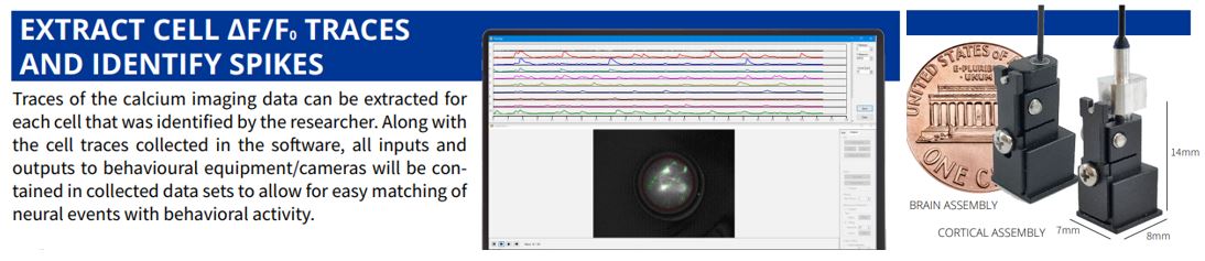
 IMAGE ACQUISITION AND ANALYSIS SOFTWARE
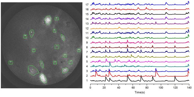
Every OASIS Implant system comes with Mightex’s Image Acquisition and Analysis software that allows users to collect large data sets of neural recordings from calcium imaging experiments and provides the key processing and analysis tools required to help users quickly convert these large data sets into results that can be clearly interpreted for analysis. The software module has been designed with the help of feedback provided by neuroscientists in the field to ensure that the platform is easy to use, integrates smoothly with typical behavioral experiments, while including the powerful advanced tools needed for studying large scale neural networks.
说到光遗传学(Optogenetics),很多英文文献的简要解释就是Controlling the Brain with Light,即通过光来控制大脑。字面理解起来很恐怖,马上联想到影帝小李子的《盗梦空间》,心有余悸啊。不过对于很多熟悉光遗传学技术的同学,脑海中肯定会浮现出以下这幅图,这是很多在体光遗传实验最常见的技术。在体光遗传技术更多的是采用光纤技术、将光源(LED或激光)引入小鼠脑区中,配合行为学研究或者电信号的记录。为什么光可以控制大脑呢?原来某些特定波长的光(如蓝光)可以打开大脑中神经细胞膜上的离子通道,使得这些神经细胞被动地变成兴奋状态,从而影响动物的某些行为和表现。这个对光敏感的离子通道(ChR2)属于一类独特的视蛋白,在蓝色光(中心波长470nm)照射的情况下,这些ChR2通道会打开,钙离子钠离子等会进入神经细胞内,引起细胞去极化产生动作电位。
以上图片描述的在体光遗传学技术的优势是设备简单、价格亲民,但局限是光纤属于盲插,光纤顶端照明的具体位置并不能精确到单个细胞的级别。对于空间精度要求比较高的光遗传学实验,越来越多课题组采用显微镜作为辅助工具,将蓝光(LED或激光)精准地并且有一定频率地照射在视野内的一群或者指定的神经细胞上。下面三幅示意图中,黑色圆圈表示整个视野,蓝色小圆圈表示蓝光照射的区域:图A是整个视野都被照射(即视野内的所有神经元都被激活);图B是只有视野中心的区域被激活;图C是视野中多个区域有选择性地被照射。满足各种光遗传学实验的需求。 那么,如果在一台已有的电生理膜片钳显微镜上,是否可以方便快捷地实现上图中三种不同的显微镜上的光遗传学技术呢?答案是肯定的。采用不同类型的LED光源或者激光器,现在已经比较成熟的解决方案,满足这些对于空间精度要求高的实验。
方案A) 采用全光谱白光LED荧光光源
传统电生理显微镜上的荧光光源采用的是汞灯,优势是光强较高,不足是汞灯灯泡寿命很短需经常更换(在屏蔽笼里更换调节很不方便),而且汞灯不能高频的开关。目前市面上的LED荧光光源稳定性及光强都能满足荧光显微镜的需求,并且LED光源可以通过外部TTL信号触发按照一定频率开关,通过放大器实现和电信号记录的完全同步。选择白光LED荧光光源需要注意以下几点:
1) 光源主机和显微镜分离,LED通过液芯光纤耦合到显微镜上,防止任何可能的电子噪声对于电信号记录的影响。这种方式也能防止光源的冷却模块产生的震动对于显微镜系统的影响。
2) LED调光旋钮可与光源主机分离,方便在屏蔽笼内直接手动调节LED光强。也可以通过USB线用软件来调节LED光强。
3) 宽光谱白光LED光源,这样可以直接替代汞灯光源,在显微镜上观察从紫外到近红外的荧光标记。同时,通过蓝色滤光片可将蓝光直接用于激活ChR2离子通道。
方案B) 采用外部光纤耦合激光器
将蓝光激光引入显微镜中,需要搭建光路、滤光片等配合,以保证激光通过物镜聚焦之后的光斑在视野中心,同时光斑大小后续可以调节。采用这个方案需要注意以下几点:
1) 激光的功率及功率稳定性都要有保证
2) 激光准直扩束之后的光斑大小需与物镜匹配
3) 选择合适的滤光片用于耦合激光
方案C) 采用DMD/DLP芯片照射多个区域
对于视野中多个区域同时照射的需求,基于数字微镜阵列技术的DMD/DLP芯片可以发挥很大的作用。类似在投影仪当中的应用,DLP芯片可以将蓝色或者黄色LED光源通过显微镜投射到样品上,单个或多个照明区域则可以通过软件设置。通过这个技术,在40倍物镜下最小照明区域可以达到0.4 x 0.4 um,实现单细胞、多细胞甚至细胞内的光刺激。采用这个方案需要注意以下几点:
1) 投影照明区域的平场均匀性,与光学成像区域匹配
2) 蓝光和黄光LED光源通过DLP芯片之后的功率
3) 投影芯片的刷新频率>1kHz,投影仪只有60Hz
4) 光刺激设备与电生理设备的同步
The
Polygon400 is Mightex’s market-leading pattern illuminator. It provides precise spatio-temporal control of light with single-cell or subcellular resolution, making it the perfect illumination tool for life science research. Compatible with any upright or inverted microscope, Polygon400 enables researchers to send light to anywhere on their specimen, and in any shape, size and complexity. In addition, multiple regions of interest can be illuminated simultaneously, and different wavelengths of light can be used with the Polygon400 for different bioscience applications. Furthermore, the Polygon400 can be seamlessly integrated via TTL into a larger system with other equipment such as electrophysiology tools or cameras.
State-of-the-Art DMD Technology
The Polygon uses digital mirror device (DMD) technology to illuminate multiple regions simultaneously. A DMD is composed of hundreds of thousands of micro-mirrors that can be individually turned on to reflect light onto the sample. Thus, you can control each mirror to control the area(s) of illumination and create any number of different sized patterns. The Polygon DMD device can be mounted into the infinity-path of any microscope.
Powerful User-Friendly Polyscan Software for Life Science Experiments
Mightex’s PolyScan software platform is bundled with every Polygon to help you execute sophisticated patterned illumination experiments for your research. It provides: Easy to use GUI to draw and define illumination patterns; Arrange sequences of illumination patterns; Define temporal illumination parameters for you experiments; Synchronize illumination patterns with other lab equipment.
The
OASIS Implant is a ground-breaking platform for simultaneous single-cell resolution optogenetics and calcium imaging in freely-behaving animals in the deep-brain, cortex, or spinal cord. Whether you are investigating a single region or multiple regions at the same time, this is the all-optical solution for your freely-behaving experiments. The OASIS Implant is purposefully designed to be reconfigurable and scalable, such that researchers can easily tackle both current and future in vivo optogenetics and imaging applications. Truly understand the link between neural circuits and behaviour using the OASIS Implant.
The OASIS Implant is not simply a product but a platform onto which you can build your complete in vivo imaging and photostimulation system. It is a reconfigurable solution that allows you to put together the components required for your current research goals including different wavelegths of illumination, spatiotemporal control of stimulation, cameras, imaging fibers, and more. By adding the necessary components to your system as time progresses, your OASIS Implant can evolve as your experiments evolve. With the OASIS Implant platform you can seamlessly transition from fiber photometry functionality to cellular-resolution calcium imaging capability or from widefield optogenetic stimulation to single-cell patterned stimulation.
IMAGE ACQUISITION AND ANALYSIS SOFTWARE
Every OASIS Implant system comes with Mightex’s Image Acquisition and Analysis software that allows users to collect large data sets of neural recordings from calcium imaging experiments and provides the key processing and analysis tools required to help users quickly convert these large data sets into results that can be clearly interpreted for analysis. The software module has been designed with the help of feedback provided by neuroscientists in the field to ensure that the platform is easy to use, integrates smoothly with typical behavioral experiments, while including the powerful advanced tools needed for studying large scale neural networks.