Scan Slide Module
The scan slide module generates a comprehensive view of specimens which exceed the conventional field of view. This is achieved by automatic scanning of a user-defined sample area and subsequent image stitching. Precise stitching algorithms assure maximal accuracy of these high resolution images.
Scanning of Multiwell Plates
The scanning technique can also be used for scanning multiwell culture plates with different sizes like 6-, 12-, 24-, 96-well formats. In addition high magnification sub-scanning within a single well is supported. To review images the standard display mode is used with easy hot-key selection of multi-stage-position, zoom and movie function.
Simultaneous Image Acquisition up to four Cameras in Multi-Camera Mode
Beside the control of multiple cameras from different vendors or models within one PC, VisiView supports up to four cameras of the same model in simultaneous mode. This allows the observation of e.g. four different fluorochromes at the same time. As a result the negative effects rela- ted to sequential image acquisition, like time delay between colors, are avoided. This function is perfectly suited for performing highly reliable ion measurements with emission ratio dyes (e.g. indo-1, cameleon) or performing co-localisation studies.
SplitView Analysis
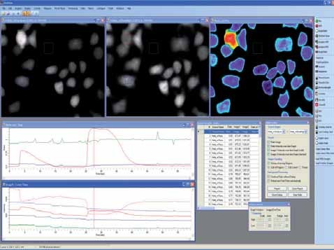
The splitview analysis allows the on-line division of images acquired with an optical image splitter, which is mounted in front of the CCD camera. This replaces time consuming post-processing and enables on-line analysis of emission ratio experiments.

Configure Well Dialog
There are different sizes of multiwell plates available. The VisiView® screening option supports 6, 12, 24, 48, 96, 384, 1536 multiwell and custom formats. For the selected plate type a plate layout is created with calibration values for offset, distance between columns and rows, well size and well shape. In addition multiple sites for each single well can be selected, e.g. 16x16 if higher objective magnification is used for high resolution imaging mode. The specimen settle time optionally prevents from taking blurred images of samples moving due to inertia.
Autofocus
The autofocus module calculates the optimal focal section for each multiwell sample in reflected-light, transmitted-light and fluorescence. For images that are recorded as time lapse or at different well positions, the cells are automatically refocused, if focus shift appears.
Object Analysis - Cell Counting
Measure or count cells automatically with a wide range of object classifiers. The object analysis tool makes it possible to determine morphometric parameters from the specimen and to report it automatically into Microsoft Excel® or text format. The well arranged object analysis dialog selectively displays filter functions, sum of object statistic or single object values. Again, VisiView's unmatched on-line functionality offers simple on-line adjustment of threshold intensity values to improve the object segmentation and the analysis results

Making Measurements
Regions of interest, circular, rectangular or polygon shape can be placed on every raw source or ratio images to display the intensity value or ion concentration.The measurements can be done simultaneously. Regi- ons can be repositioned using the history function if the cell has moved or the stage was touched. Individual region position can be frozen or changed by editing the current region or even all regions.
Trigger Protocol
For flexible experiment control, complex user defined trigger sequen- ces can be important. The trigger protocol option offers variable on/off switching of external devices during all kinds of experiment sequences.
